
Ronen Nazarian, M.D.
Otology,
Neurotology & Hearing Surgeon

Hootan Zandifar, M.D., F.A.C.S
Facial Plastic and Reconstructive Surgeon
Deformities of the auricle can be divided in two categories. They can be either external ear malformations or external ear deformities.
An external ear malformation is described as an ear that is missing part or all of the external ear cartilages. This is usually named Microtia and requires surgical correction to restore the normal shape of the external ear. Often times, Microtia is associated with hearing issues and an evaluation by an Otologist (Fellowship trained ear surgeon) is recommended to see if natural hearing can be restored.
An external ear deformity, on the other hand, is characterized by fully formed ear cartilages that are abnormally shaped. These deformities are typically not associated with hearing loss, however, an evaluation with an Otologist (A fellowship trained ear surgeon) is ideal to rule out any internal deformities. There are several different types of external ear deformities. They can include:
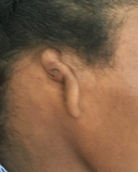
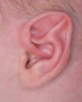
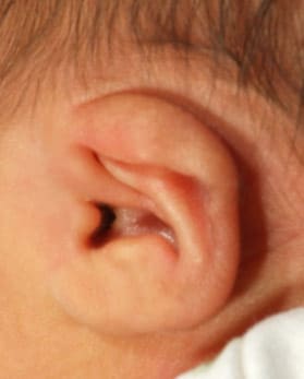
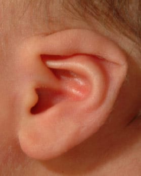
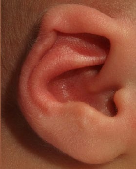
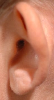
Correction of above external ear deformities can take place in either of the two different times of life. First (and most preferable) time that these deformities can be corrected is within the first two weeks of age. Because maternal hormones are still circulating inside the infant’s blood, the ear cartilage during these first weeks of life is still pliable and moldable. Thus ear molding can be performed during this time with excellent cosmetic outcomes. Ear molding can correct over 95 percent of all pediatric ear deformities.
EarWell™ Infant Ear Correction System makes the process of ear molding easier on the parents and infants. Ear molding treatment typically lasts anywhere from 2-6 weeks and should be started by the 2nd week of life for the best results. The treatment is painless, incision less and requires no anesthesia or numbing. There is no recovery time needed after this treatment.
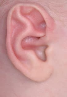
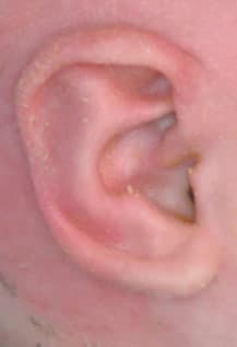
If the correction for an external ear deformity is not performed during the first few weeks of life, the next time period that a correction can be done is usually after the age of 5. By then the ear has reached adult size and surgical correction will not interfere growth of the cartilage. The surgical correction at this stage is called otoplasty.
Otoplasty is typically done for protruding ears, however, any of the above-mentioned ear deformities can be addressed via otoplasty. During this surgery excess cartilage can be removed or abnormal cartilage can be re-shaped to look like normal ear. Otoplasty is typically done via a well-hidden incision behind the ears and has a relatively short recovery time. Anesthesia is usually required for this surgical procedure. Although otoplasty can be performed anytime after the age of 5, it is usually recommended to obtain correction of the ear deformity prior to start of the school. This will minimize the psychological trauma that is usually incurred from teasing by other children.


Microtia is a relatively common congenital ear deformity where the ear is either underdeveloped or completely absent (also referred to as anotia). It can be associated with a narrow ear canal or no ear canal at all. Depending on the degree of the deformity, various surgical techniques can be used to reconstruct the ear. Microtia is usually classified on the degree of ear development. The most common classification is grade III microtia. In this grade most of the external ear or pinna is not formed and there is no ear canal present.
Grades of Microtia
- Grade I: Slight underdevelopment of the external ear and external ear canal. Normal ear structure and landmarks are preserved.
- Grade II: Partially undeveloped external ear with absence of classic ear structure and landmarks. The external ear canal is typically sealed/obstructed. Middle ear structures such as the ear drum are present. Conductive hearing loss is typical.
- Grade III: Near total absence of identifiable external ear structure, external ear canal, and ear drum.
- Grade IV: Complete absence of the external ear, external ear canal, and ear drum.
Treatment
Several surgical techniques have been used to reconstruct the deformed ear:
- One technique uses cartilage, typically harvested from the ribs to reconstruct the shape of an ear. This is then implanted in the skin behind the malformed ear and raised, in stages, to look more like a normal ear.
- Another technique uses an implantable material such as Medpore® that is already shaped to resemble an ear. This is provided with blood supply and covered with the patient’s own skin. This technique usually requires one stage.
The absence of an ear canal leads to a type of hearing loss referred to as conductive hearing loss. Restoration of hearing can be accomplished by either of the following methods:
- Implantable Hearing Aid: An implantable hearing aid can be used to transmit the sound waves through the bones of the skull and restore some of the hearing.
- Reconstruction: In some patients, the ear canal can be re-constructed to restore natural hearing without the use of hearing aids.
- The procedures above are typically performed after the patient is at least 6 years of age to ensure that the other ear has fully grown to adult size for comparison.
When patients are considering treatment, it is important to select a surgical team that has both a fellowship-trained facial plastic surgeon and an otologist (ear specialist). This will ensure that you not only achieve the best cosmetic results, but also reach your highest hearing potential.


